
A new scaffolding technique developed by researchers at Lund University in Sweden offers a promising advancement in bone repair, potentially revolutionizing the treatment of significant skeletal injuries. The innovation centers around a cell-free cartilage support structure designed to guide the body’s natural bone regeneration processes.
The technique involves removing living cells from lab-grown cartilage tissue whereas preserving its essential biological structure and signaling cues that promote cell growth. This creates a biocompatible framework that directs bone repair, allowing the body to rebuild damaged tissue step-by-step, according to Maspero.
Early trials in animal models have shown encouraging results, with the new scaffold successfully stimulating bone growth without triggering strong immune responses. Researchers are now preparing to move forward with clinical trials in humans.
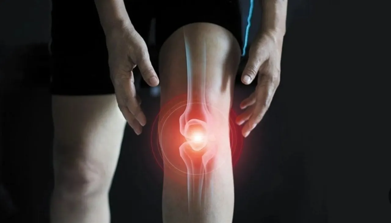
Bone and skeletal injuries are a leading cause of long-term disability worldwide. In many cases, such as the removal of cancerous tumors or severe joint diseases like rheumatoid arthritis and osteoarthritis, or serious infections, the body is unable to repair bone on its own. These situations often require bone grafting procedures to restore structure and function. More than 2 million people globally require bone grafting each year, researchers estimate.
Current treatments often rely on using tissue or cells from the patient’s own body, a process that can be costly, time-consuming, and physically demanding. The new technique aims to address these challenges.
“Current treatments designed for each patient individually are not always optimal,” explained Alejandro Garcia, a specialist in skeletal molecular biology at Lund University. “Patient-specific grafts are expensive and time-consuming, and they don’t always succeed. A more universal approach to tissue engineering, based on manufacturing reproducible materials, offers significant advantages.”
The development of the technology began with the team cultivating cartilage tissue in the laboratory. They then removed all living cells through a process known as decellularization. While the cells were removed, the extracellular matrix – the network that gives tissues their shape and contains growth signals – remained intact. When this structure is implanted at the site of injury, it acts as a natural template, guiding the body’s cells to build new bone.
Paul Borgin, a professor of skeletal molecular biology and the study’s lead researcher, suggested the new technique could pave the way for the production of ready-to-use bone grafts. “The material we have developed is based on stable and reproducible cell lines and can stimulate bone formation without triggering strong immune responses,” Borgin said. “We have demonstrated the possibility of developing an off-the-shelf graft that can repair large bone defects.” He added that the ability to produce this material in advance and store it represents an important step toward its future use in clinical practice.
A key advantage of this technology is that it doesn’t require a custom design for each patient, meaning it has the potential for widespread use. The research team is now focused on identifying the types of injuries that will be tested first in human studies, such as severe defects in the long bones of the limbs, alongside completing the necessary regulatory and ethical procedures to launch clinical trials. They are also working on developing a manufacturing system capable of producing the scaffold on a large scale while maintaining high standards of quality and safety.