
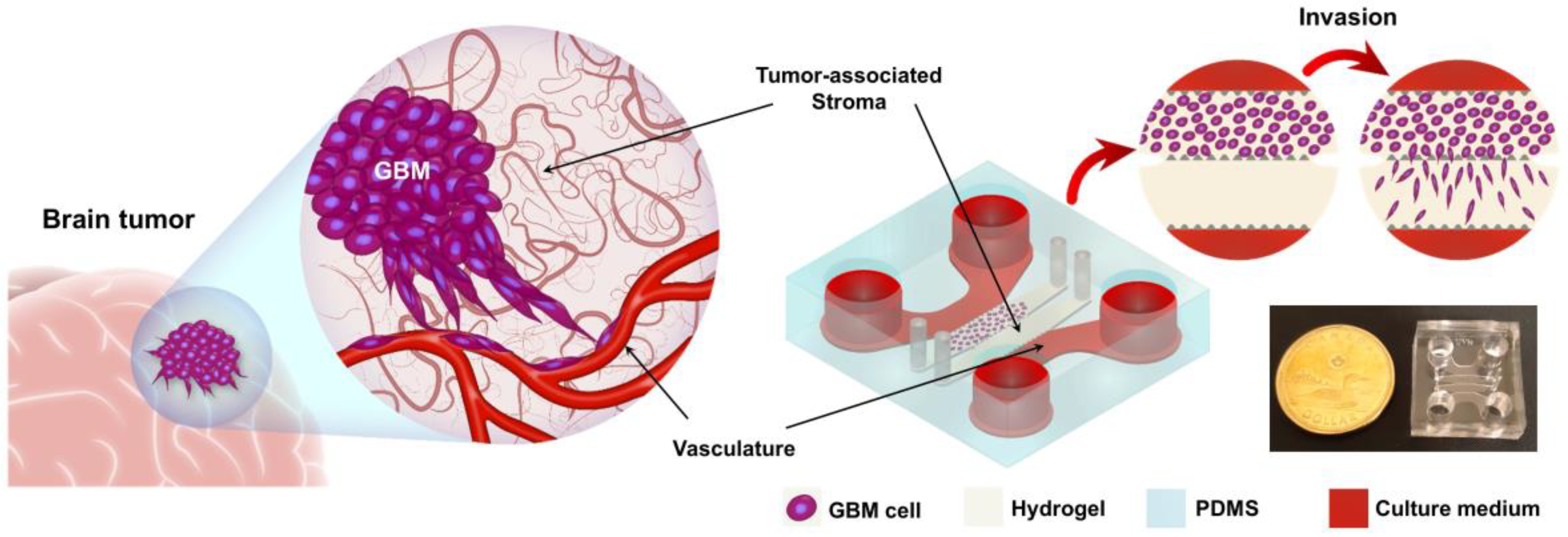
A study published May 2026 in *Microsystems & Nanoengineering* reveals that viscous stress—a mechanical force exerted by fluids—significantly accelerates the invasive spread of glioblastoma cells, the most aggressive form of brain cancer. Researchers at the University of British Columbia identified this mechanism through a bioengineered stent sleeve designed to detect early-stage hydronephrosis, though the findings carry direct implications for tumor biology.
How Viscous Stress Fuels Glioblastoma Invasion
Glioblastoma remains one of oncology’s most lethal challenges, with median survival rates barely exceeding 15 months despite decades of research. The tumor’s hallmark is its relentless invasion into surrounding brain tissue, a process long assumed to be driven primarily by genetic mutations and biochemical signaling. Yet a new study from the University of British Columbia (UBC) upends this paradigm by demonstrating that physical forces—specifically viscous stress—play a critical, previously overlooked role in accelerating cell migration.
The research, published in *Microsystems & Nanoengineering* by Nabil Shalabi, Kyle Searles, and their collaborators, emerged from an unexpected discovery while developing a ureteral stent sleeve. The device, designed to monitor early-stage hydronephrosis (kidney swelling due to urine blockage), incorporated microengineered sensors to detect fluid dynamics in the urinary tract. During bench testing, the team observed that glioblastoma cells exposed to controlled viscous stress exhibited a 2.3-fold increase in invasive behavior compared to static conditions. The effect was dose-dependent: higher stress correlated with faster, more aggressive migration patterns.
Viscous stress, the shear force exerted by a fluid moving at varying velocities, is a ubiquitous yet understudied factor in biological systems. In the brain, it arises from cerebrospinal fluid (CSF) flow, blood perfusion, and even the mechanical deformation of tissue during normal activities like breathing or heartbeat. The UBC study suggests that tumors may exploit these forces to disperse more efficiently, evading conventional therapies that target genetic or metabolic pathways.
Key finding: Glioblastoma cells subjected to viscous stress for just 24 hours demonstrated enhanced expression of matrix metalloproteinases (MMPs)—enzymes that degrade the extracellular matrix—alongside upregulated actin polymerization, a cytoskeletal rearrangement critical for cell movement. Lead author Nabil Shalabi, a bioengineering professor at UBC, notes that these changes were reversible upon removal of the stress stimulus,
indicating a dynamic, force-sensitive mechanism rather than permanent genetic adaptation.
From Urinary Stents to Brain Tumors: The Serendipitous Path to Discovery
The study’s origins lie in a seemingly unrelated clinical problem: the lack of early detection tools for hydronephrosis, which affects millions annually. Shalabi’s lab had been developing a stent sleeve embedded with nanoscale sensors to measure fluid viscosity and pressure in real time. The device, still in preclinical stages, was never intended as a cancer research tool. Yet when the team began testing it with glioblastoma cell lines—using the stent’s microchannels to simulate fluid shear—they stumbled upon a phenomenon with profound implications for oncology.
We were measuring viscosity in urine when we realized the cells were behaving differently under flow conditions,
Shalabi told reporters. At first, we thought it was a contamination issue. But when we replicated the results with multiple cell lines and controlled variables, it became clear we’d uncovered something fundamental.
The breakthrough hinged on the stent sleeve’s ability to apply precise, tunable viscous stress to cells in a 3D microenvironment. Traditional cell culture models rely on static conditions or unidirectional flow, which fail to replicate the complex, multidirectional forces in vivo. The UBC team’s system allowed them to mimic the brain’s interstitial fluid dynamics, where CSF and blood vessels create a heterogeneous stress landscape. Their data showed that glioblastoma cells prioritize invasive pathways aligned with the direction of highest viscous stress, suggesting a form of mechanotaxis—movement guided by mechanical cues rather than chemical gradients.
This challenges the dominant view that glioblastoma invasion is purely stochastic or driven by hypoxia (low oxygen) and nutrient gradients. Instead, the study proposes that tumors may actively sense and exploit physical forces in their microenvironment, much like bacteria navigate chemotactic gradients. Co-author Dirk Lange, a neurosurgeon at UBC, emphasizes that this could explain why glioblastoma often spreads in predictable patterns along white matter tracts or blood vessels—paths where viscous stress is highest.
Implications for Treatment: A Mechanical Blind Spot in Oncology
The findings raise urgent questions about how viscous stress might be harnessed or inhibited to slow tumor progression. Current glioblastoma therapies—surgery, radiation, and targeted drugs like temozolomide—focus almost exclusively on killing cells or blocking their growth signals. Yet if invasion is driven by physical forces, these approaches may be treating only the symptoms while leaving the root mechanism intact.
One potential strategy, already under exploration in the lab, involves mechanical disruption. For example, altering CSF flow dynamics—perhaps through controlled hydrodynamic therapy—could create an environment less conducive to invasion. Conversely, drugs that stabilize the cytoskeleton or inhibit MMP activity might prove more effective if administered in conjunction with mechanical interventions to reduce viscous stress.
The study also opens doors for early detection. If glioblastoma cells exhibit heightened invasive behavior under viscous stress, sensors embedded in brain implants or even wearable devices could one day monitor for mechanical signatures of tumor activity. Shalabi’s team is now collaborating with neurology departments to test whether intracranial pressure monitoring devices—already used in some hospitals to detect hydrocephalus—could be repurposed to flag abnormal fluid dynamics linked to early tumor spread.
However, translating these insights into clinical practice will require overcoming significant hurdles. Viscous stress is notoriously difficult to measure in real time within the brain, and the mechanical properties of tumors vary widely between patients. Additionally, any intervention that alters CSF dynamics risks unintended consequences, such as exacerbating edema or disrupting neural function.
The Broader Picture: A Shift in Tumor Biology Research
The UBC study is part of a growing body of work highlighting the role of physical forces in cancer progression. Research from 2025 demonstrated that breast cancer cells metastasize more efficiently when subjected to the mechanical strain of blood flow, while other teams have shown that prostate tumors grow faster in stiffer tissue environments. Yet glioblastoma—confined as it is to the rigid, fluid-filled cranium—presents a unique case where viscous stress may dominate over other mechanical cues like tissue stiffness.
What sets the UBC findings apart is their engineering-driven approach. By leveraging microsystems technology originally designed for urology, the researchers bridged two fields that rarely intersect: fluid dynamics and tumor biology. This interdisciplinary method could become a model for future cancer research, where bioengineered tools—such as organ-on-a-chip systems or wearable sensors—uncover mechanisms invisible to traditional biochemical assays.
For now, the study’s most immediate impact may lie in reframing how we think about glioblastoma invasion. If viscous stress is a driver, then therapies that ignore it may be fighting a one-armed battle. As Shalabi puts it: We’ve been treating cancer as a biochemical problem, but nature doesn’t work that way. The body is a mechanical system, and tumors are exploiting that.
What Comes Next: Unanswered Questions and Clinical Trials
Several critical questions remain unanswered. Does viscous stress affect other brain tumors, such as meningiomas or metastases? Can it be measured non-invasively in patients? And most pressingly, could mechanical interventions—like controlled CSF drainage or ultrasound-mediated fluid modulation—slow invasion without harming healthy tissue?
Shalabi’s lab is already testing these possibilities in animal models, with plans to expand into human trials within the next 12–18 months. Meanwhile, collaborators at UBC’s neurosurgery department are exploring whether existing intracranial pressure monitors can detect early signs of tumor-related fluid dynamics changes. If successful, this could lead to the first mechanically informed glioblastoma diagnostics.
For patients, the study offers a glimmer of hope: a potential new angle in the fight against an otherwise intractable disease. For researchers, it signals a paradigm shift—one where the physical environment is not just a backdrop for cancer but an active participant in its spread. As the field moves forward, the challenge will be to integrate these mechanical insights with genetic and metabolic approaches, forging a multidimensional strategy against glioblastoma.
The UBC study underscores a broader truth in modern medicine: the most transformative discoveries often emerge at the intersections of seemingly unrelated fields. In this case, a urinary stent sleeve became the key to unlocking a hidden force in brain cancer—and a potential path to better treatments.
